

SCAN IN. REPORT OUT.
Meet OmniBioFex 1.0 47B — a combined vision + text engine (27B vision head, 20B text head) that reads a scan and writes a structured clinical report: findings, impression, differential diagnosis, and management. Four scan modes, four languages, six templates.
Four Scan Modes
Every scan is a choice. Pick the mode that matches the question you're asking. Prices are debited from your wallet per successful run — failures auto-refund.
STANDARD
The full report — findings with confidence, impression, advice, differential, and management. Optional 2-pass localization verification.
→ RECOMMENDED FOR ROUTINE READSMULTI-MODEL
Two independent vision passes on the same image. Agreement scores per finding. Best for ambiguous studies or high-stakes reads.
→ TWO PASSES · AGREEMENT SCOREFIND ABNORMALITIES
Sweeps the study for fractures, opacities, and lesions. Returns bounding boxes with severity colour-coding on the original image.
→ ANNOTATED BOUNDING BOXESCOMPARE
Upload a prior and a current study. The engine returns interval changes, new findings, and resolved findings side-by-side.
→ PRIOR + CURRENT · INTERVAL REPORTWatch It Think
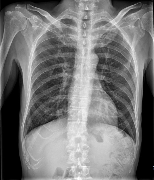
A live replay of what a scan actually goes through. The beam sweeps the frame, the log streams the pipeline's real stages on a real study.
Medical Privacy Mode
A toggle inside the app that turns the terminal into a shared-room-safe workstation. One click. Everything else in the product keeps working exactly the same.
What You Get
Every scan produces a formal clinical imaging report. Structured like a radiologist's report, exportable to a multi-page PDF, with a live follow-up chat that never loses context.
EXAM TITLE + VIEW
Auto-detected — "CHEST X-RAY · PA View", "CT BRAIN · Axial", "MRI KNEE · Sagittal".
STRUCTURED FINDINGS
6–12 discrete clinical sentences. Each with confidence and a 3×3 region tag.
IMPRESSION
One-sentence diagnostic conclusion. "No abnormality detected" when normal, or the leading diagnosis otherwise.
CLINICAL ADVICE
One-line recommendation — often "clinical correlation recommended" or a specific next step.
DIFFERENTIAL DIAGNOSIS
3–6 conditions ranked most-to-least likely, each with a short rationale.
MANAGEMENT
3–6 specific next steps — imaging, labs, referrals, or conservative management.
PDF · MD · JSON EXPORT
Multi-page PDF with letterhead. Markdown for docs. Raw JSON for pipelines.
FOLLOW-UP CHAT
Ask anything about the report — image-grounded, zoom-aware. $0.01 per turn. Saved forever.
How It Works
Sign in with Google. Attach a scan. Enter the patient's name, age, gender, language, and template (all optional except the image). That's it — the pipeline does the rest in 15–40 seconds depending on mode.
SIGN IN
One-click Google OAuth. No passwords, no email verification, no credit card. Account live the moment you consent.
ATTACH SCAN
Drop a chest X-ray, CT, MRI, or histopathology slide. Full image, no cropping. DICOM is converted in-browser. Client-side compression to 1600px.
PICK MODE + DETAILS
Standard, Multi-model, Find, or Compare. Optionally add patient name, age, sex, language (4), and template (6).
ENGINE RUNS
Four sequential steps: vision extraction (27B) → impression → differential + management (20B) → final assembly.
REPORT + FOLLOW-UP
The report appears in chat. Click EXPORT PDF for a clinical-grade document. Then ask anything — follow-up chat is live at $0.01/turn.
What the Chat Handles
The chat is not a form. It is a live session. Every message is saved, every image is preserved, and follow-up questions are grounded in the original study.
MULTI-IMAGE UPLOAD
Up to 2 images per request in Compare mode. Single image in all others.
DICOM SUPPORT
Upload a .dcm file and the browser converts it to a JPEG before sending. Single-frame only.
FOLLOW-UP Q&A
Ask anything about the report. Image-grounded. Zoom-aware. $0.01 per turn.
EXPORT ANY TIME
PDF · Markdown · JSON — from any report, at any point in the chat.
PERSISTENT HISTORY
Every session is saved. Reload the page — it's all still there.
4 LANGUAGES · 6 TEMPLATES
English, Hindi, Tamil, Telugu. General, Chest, Neuro, Ortho, Cardiac, Patient-Friendly.
DARK MODE
One toggle. Light for clinical review, dark for reading rooms.
MEDICAL PRIVACY MODE
Redacted previews and 15-min idle signout when you share the terminal.
Stage-by-Stage
Click any stage. Watch how a single image travels through the pipeline and becomes a structured report.
Live Terminal
A real-time peek at what a running inference actually looks like on the backend.
Why Two Heads?
Vision and reasoning are different problems. A single monolithic model is a compromise on both. So we split them.
READS THE IMAGE
Extracts 6–12 discrete findings with per-item confidence and 3×3 region tags. Runs a parallel fracture-hunt audit. Emits strict JSON.
WRITES THE REPORT
Takes the structured findings, writes the one-line impression, ranks a differential diagnosis, and drafts 3–6 management steps.
ROUTED, NOT STACKED
Each stage calls the head that is best for it. Routing beats stacking — predictable memory, predictable cost.
Built for Teams
Owners on the Enterprise tier can top up a single shared wallet and invite up to 10 seats. Every seat shares the same pipeline; every action is audited.
UP TO 10 MEMBERS
One wallet, one shared history. Owner invites by email, sets role, transfers seats between members.
5 ROLES
Admin, Member, Technician, Reviewer, Billing — each with scoped permissions inside the team panel.
SHARED TEAM WALLET
Owner tops up once; every member scans from the same balance. Every member can switch between personal and team wallet with one click.
WHITE-LABEL PDF
Upload a clinic name and logo (PNG/JPEG/WebP ≤ 500 KB). It appears on every team member's PDF letterhead.
FHIR WEBHOOK
Completed reports POST as HL7 FHIR DiagnosticReport to any HTTPS endpoint. Save + send test from the team panel.
AUDIT LOG + SPEND LIMIT
Every action is logged. Set an optional monthly USD ceiling on team-wallet spend.
DEVELOPER API KEY
One key per wallet. POST to api.omnibiofex.cloud/apiInference with Authorization: Bearer. Same model, same rate.
PREPAID, NO SURPRISES
No subscriptions. No auto-renewal. Failed calls auto-refund. Balance never expires.
Specification Sheet
OmniBioFex 1.0 47B is a routed combination of two model heads served together on Groq's LPU stack. A 27B vision head reads the image. A 20B text head writes the report. Combined: 47B parameters — chained, not stacked.
Benchmarks
Benchmark figures are the published numbers for the underlying vision head (qwen/qwen3.8-27b). They describe the model, not the combined pipeline.
Capabilities
One engine. One report format. All output is decision-support material for qualified professionals.
01 · CHEST X-RAY
Lung fields, cardiac silhouette, mediastinum, pleural spaces, osseous structures.
02 · CT — SINGLE FRAME
Descriptive observations on single CT slices. The model does not reconstruct 3D volumes.
03 · MRI — SINGLE FRAME
Descriptive observations on single MRI frames. Not a substitute for a radiologist's read.
04 · MUSCULOSKELETAL
Radiograph observations. Not a fracture-detection product — findings require professional review.
05 · TISSUE IMAGING
Tissue images. Not a pathology AI; output must be reviewed by a pathologist.
06 · SKIN IMAGING
Skin lesion images. Output must be reviewed by a dermatologist before clinical use.
Ready to start?
Free tier, or top up a wallet and skip the cooldown. No card stored. No auto-renewal.